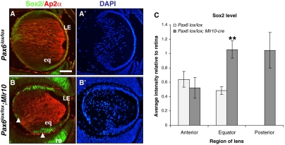
Fig. 6.
Pax6 downregulates Sox2 in the lens equator. (A,B) Immunofluorescent detection of Sox2 (green) and Ap2α (red) in E14.5 control (A) and Pax6lox/lox;Mlr10 (B) mouse lenses. Arrowheads in B indicate elevated expression of Sox2 at the lens equator and in the posterior lens. (A′,B′) Counterstaining of A,B with DAPI. (C) Quantification of Sox2 protein by confocal image analysis (n=6, **P<0.001). eq, lens equator; LE, lens epithelium; re, retina. Scale bar: 100 μm.

