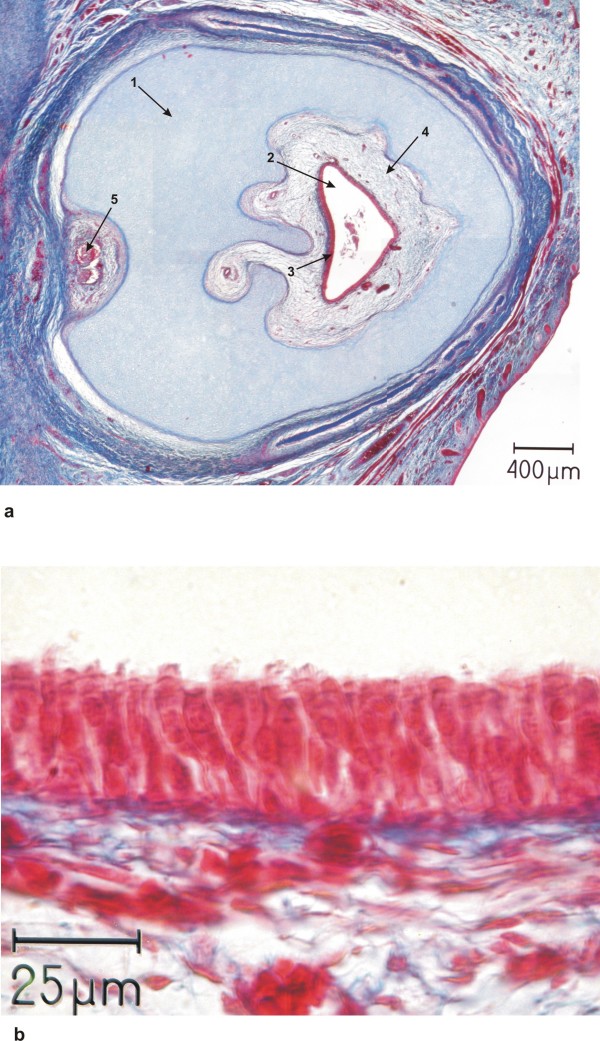
Figure 9.
Histology of proboscis. a) Overview of a histological section of the proboscis showing the closed circular cartilaginous wall (arrow 1) with a central canal (arrow 2) with an epithelial lining (arrow 3) supported by loose connective tissue (arrow 4) which contains blood vessels and two nerves at the posterior side (arrow 5). b) Higher magnification of the epithelial lining of the central canal, depicting a respiratory epithelium with cilia (arrow 1) and goblet cells (arrow 2).

