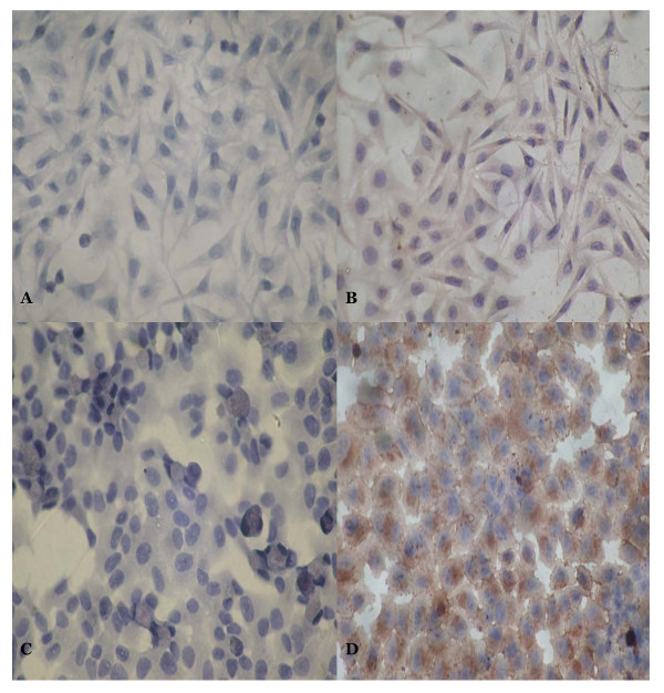
Figure 4.
The immunohistochemistry results of FBG2 in MKN-PC, MKN-FBG2, HFE-PC and HFE-FBG2 cell lines. A: There was no positive signal in MKN-PC cell. B: There was positive signal in MKN-FBG2 cell. The brown positive signals were mainly distributed in cytoplasm. C: There was no brown positive signal in HFE-PC cell too. D: There was positive signal in HFE-FBG2 cell and the brown positive signals were mainly distributed in cytoplasm and cell membrane. The results showed that there were expressions of FBG2 gene in MKN-FBG2 and HFE-FBG2 cell lines. (×200)

