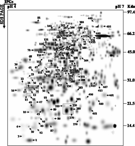
Fig. 4.
Higher level match set of protein spots detected by 2-D electrophoresis. The match set was created in silico from seven standard gels for each of the time points as depicted in supplemental Fig. 1. For the sake of clarity the protein spots within the dashed frames represent the enlarged sections in Fig. 5. The numbers indicate protein spots listed in supplemental Table 2 that were significantly identified by MS/MS analysis.

