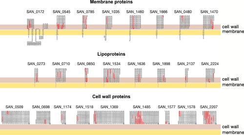
Fig. 2.
Schematic topological representation of each protein identified on the surface of COH1 GBS strain. Topological organization of the identified proteins was predicted using PSORTb information. The orange line represents the membrane, and the red line represents the cell wall. For lipoproteins, the lipoyl anchor is represented as a black segment embedded within the membrane, whereas for the cell wall-anchored proteins, the last C-terminal blue residues are the LPXTG signature anchored to the cell wall. The identified peptides are marked in red.

