
Figure 1.
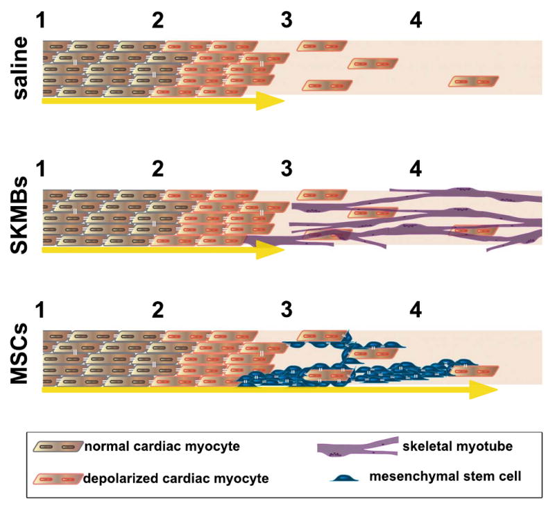
(A) Following the infarct, cells in the border zone (between 2 and 3) are depolarized, and the few surviving myocytes in the infarct zone (between 3 and 4) are poorly coupled to the border, prohibiting electric current flow into this region (yellow arrow depicts current flow terminating at interface between border zone and infarct). (B) When skeletal myoblasts (SKMBs) are added to the infarct zone they do not electrically couple, and thus do not promote electrical function in the infarct. (C) Mesenchymal stem cells (MSCs) couple to the surviving myocytes in the border and infarct zones, permitting electrical current spread into this region (depicted by yellow arrow extending into infarct zone). They may also enhance survival via angiogenesis or promote myocyte proliferation in the infarct by secretion of paracrine factors. The cartoon describes a localized region and is intended only to illustrate the basis of enhanced electrical conduction, not the diffuse (MSC) or punctuate (SKMB) distribution of the delivered cells as proposed by Mills et al.