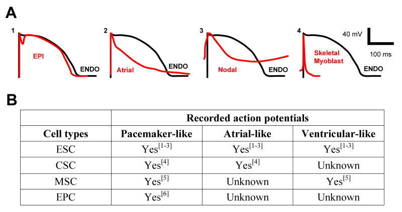
Figure 2.
(A) Differences in times of repolarization will result in electrical dispersion. (1). The differences in the action potentials (APs) across the ventricular wall—between epicardial (EPI) and endocardial myocytes (ENDO)—result in normal ventricular dispersion. (2) If an atrial myocyte is implanted into the ventricle, its action potential creates enhanced dispersion of repolarization compared with ventricular endocardial myocytes. (3) Enhanced dispersion of repolarization will occur when cells expressing nodal potentials are implanted into ventricle. (4) Even if skeletal myoblast are engineered to express connexins, the dispersion of repolarization between these cells and ventricular myocytes would be greatest of all the cell types shown. Schematics of action potentials adapted from the following references: Nodal AP from [23]; EPI and ENDO from [24]; Atrial AP from [25]; Skeletal myoblast from [26]. (B) When induced to differentiate, all of the cell types currently under investigation for cardiac repair yield heterogenous populations of cardiac myocytes. Delivery of such mixtures of cells could result in the type of electrical dispersion described above and induce fatal arrhythmias. (ESC = embryonic stem cell; CSC = cardiac stem cell; MSC = mesenchymal stem cell; EPC = endothelial progenitor cell).

