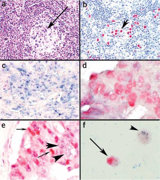
Figure 2.
Determination of putative targets of miRNAs by colocalization analysis with immunohistochemistry. The miRNA miR-155 is markedly upregulated in lymphoproliferative diseases, such as AIDS, and is also increased in colon cancer in which there is often dysregulation of the DNA repair gene MSH 2. (a) miR-155 is present in the atypical lymphocytes but not the endothelial cells as determined by in situ hybridization with an LNA probe; the arrow points to the germinal center where several blue-staining cells indicative of miR-155 expression are seen. Many more miR-155 cells are seen in the interfollicular zone. (b) By analyzing serial sections with immunohistochemistry using an antibody directed against HHV8 and a red chromogen, it was simple to demonstrate that there were over 25 times more miR-155-positive cells than HHV8-positive cells, showing that HHV-8 per se was not responsible for upregulating miR-155; the arrow points to several HHV-8 red staining cells. (c) We performed colocalization experiments with miR-155 and MSH 2, using cell conditioning for 30 min for the MSH2 protein. Note that in cancer cell groups strongly positive for miR-155 (blue, in situ hybridization with an LNA probe), MSH 2 (red, immunohistochemistry) is not evident. (d) Conversely, in colon cancer cell groups where the neoplastic cells expressed MSH 2 (red, immunohistochemistry), miR-155 (blue, in situ hybridization with an LNA probe) was not evident. (e) In some groups of cancer cells, both miR-155 and MSH 2 were present. The colocalization experiments in these groups showed that the cells making MSH 2 (small arrows, red—immunohistochemistry) did not contain miR-155 (large arrows, blue—in situ hybridization with an LNA probe) and vice versa. These data suggest that miR-155 may be downregulating MSH 2. (f) Colocalization experiments with cell lines can also be used to get useful information on whether a given miRNA may be regulating a specific protein. miR-16 downregulates the protein bcl-2, which is an important anti-apoptotic factor in several lymphomas. Colocalization experiments showed that miR-16-positive cells (small arrow, blue—in situ hybridization with an LNA probe) were bcl-2 negative, and the bcl-2-positive cells (large arrow, red—immunohistochemistry, ) were miR-16 negative, lending supporting evidence to a direct role for miR-16 in bcl-2 regulation. Panels a–c are at ×400 and panels d–f are at ×1,000 original magnification.

