Figure 1.

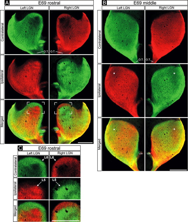
Retinogeniculate projections to the dLGN on E69. A-C, Contralateral retinal axons (top rows), ipsilateral retinal axons (middle rows), and their merged representation (bottom rows) in right and left dLGNs of an E69 macaque. A, C, Rostral E69 dLGN. B, Middle E69 dLGN. A, B, Axons from the two eyes intermingle at E69. B, Within the middle dLGN, alternating regions containing denser inputs from one eye or the other are present. These occur at mirror-symmetric locations in the two dLGNs (B, compare asterisks in middle and bottom rows). C, In the dorsal portion of the rostral E69 dLGN (see framed regions in A for orientation), eye-specific segregation is nearly complete; layer 6 (L6) receives axons from the contralateral eye, and the region directly ventral to it (L5) receives axons from the ipsilateral eye. A-C, At all locations, strong mirror symmetry is seen in the two hemispheres. Coronal plane is shown. Dorsal is up. O.T., Optic tract; LGv, ventral lateral geniculate nucleus. Scale bars, 550 μm.
