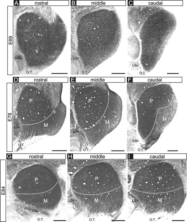
Figure 2.

Cytoarchitectural differentiation of the dLGN. A-I, Photomicrographs of thionin-stained tissue sections of the embryonic macaque dLGN at E69 (A-C), E78 (D-F), and E84 (G-I). Rostral (A, D, G), middle (B, E, H), and caudal (C, F, I) levels are shown for each age. At the ages shown here, there are no intralaminar spaces corresponding to eye-specific cellular layers, consistent with previous reports (Rakic, 1977a). A-C, At E69, the M and P divisions cannot reliably be distinguished at all rostrocaudal locations. However, in the E78 (D-F) and the E84 (G-I) specimens, the M and P divisions can be broadly distinguished on the basis of cell spacing and morphology of the dLGN at all rostral, middle, and caudal locations. Dotted lines indicate the boundaries between the M and P divisions. Coronal plane is shown. Dorsal is up, and medial is to the left. O.T., Optic tract; LGv, ventral lateral geniculate nucleus. Scale bars, 500 μm.
