
Figure 5.
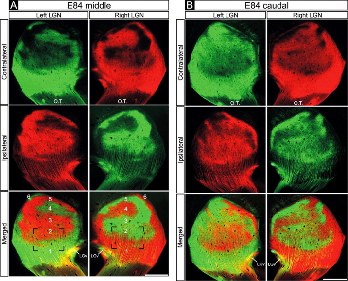
Retinogeniculate projections to the dLGN on E84. A, B, Contralateral retinal axons (top rows), ipsilateral retinal axons (middle rows), and their merged representation (bottom rows) in the right and left dLGNs of an E84 macaque. A, Middle dLGN. B, Caudal dLGN. A, B, Eye-specific segregation is present throughout the dLGN. A, All six eye-specific domains (1-6) are visible. A, B, Note the high degree of mirror symmetry in the pattern of eye-specific inputs in the two dLGNs. Coronal plane is shown. Dorsal is up. O.T., Optic tract; LGv, ventral lateral geniculate nucleus. Scale bars, 550 μm.