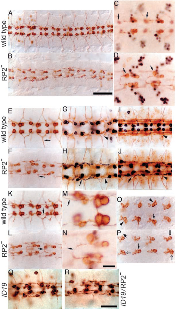
Fig. 3.
Without Eve, RP2 and a/pCC neurons show abnormal axonal morphologies. CNS preparations from embryos carrying both transgenes UAS-τlacZ (microtubule-associated βgal marker) and RN2-Gal4 (RP2+a/pCC driver), in a wild-type background (A,C,E,G,I,K,M,O), a ARP2A mutant background (eve null rescued with RP2 element-deleted transgenes; B,D,F,H,J,L,N,P), in an eveID19 background (Q) or in eveID19, ΔRP2A transheterozygotes (R), as indicated beside each row. (A,B) Anti-β-gal staining; overview of the CNS. Scale bar (in B): 50 µm. (C,D) Anti-Eve staining (black) followed by anti-β-gal staining (brown); black arrows indicate RP2 neurons. The focal plane is that of the U/CQ neurons, so that the RP2s are slightly out of focus. Note that RP2 is abnormally close to aCC in the mutant. (E,F) Anti-β-gal staining; higher magnification view of A,B in the RP2 and aCC axonal focal plane. Note that very few RP2 axons turn laterally (arrows) in the mutant. (G–J) Anti-β-gal staining (black) followed by anti-Fas2 staining (1D4 antibody, brown); stage 13 (G,H) and stage 15 (I,J) are shown. In the mutant, RP2s often extend an axon posteriorly, rather than anteriorly as in the wild type, along the lateral longitudinal fascicle (arrow in H,J). Although the majority of RP2s extend an axon anteriorly, which then either turns laterally at the pISN (arrowhead in J), as in the wild type, or fails to turn at the ISN (arrowhead in H; compare with the wild type in G,I), most of them do not exit the CNS (see Table 1). Even those that do exit the CNS fail to extend to the dorsal muscle field (see Fig. 5). (K,L) Anti-β-gal staining; higher magnification view of A, B in the pCC axonal focal plane. The pCC axons extend anteriorly beyond the next more anterior pCC cell body in the wild type, while in the mutant, the pCC axons often cross the midline at the anterior commissure (arrows). Note that there are small neurons extending their axons laterally in the wild type. These are RP2 siblings, because at earlier stages, they also stain for Eve (not shown). (M,N) Higher magnification of K and D, respectively. Scale bar (in N): 5 µm. (O,P) Anti-β-gal staining; stage 12 CNSs are shown. In the wild type, the positions of the aCC and pCC cell bodies (after their generation from GMC1-1a) are well regulated; pCC is positioned either posteriorly (arrow) or posteriorly and laterally (arrowheads) relative to aCC. This positioning is disarrayed in the mutant; pCCs positioned posteromedially (wide arrow) or directly laterally (open arrows) are indicated. (Q,R) Anti-β-gal staining; the temperature-sensitive eve allele ID19 kept at the restrictive temperature during nervous system development after allowing segmentation to occur at the permissive temperature (see Materials and methods for details). (Q) eveID19 homozygous mutant; note that many more axons extend laterally than in ΔRP2A/ΔRP2A (compare with F), indicating that eveID19 does not act as a complete null allele in the nervous system. (R) A single copy of eveID19 with one copy of ΔRP2A; note that the phenotype is more severe than that of eveID19 homozygotes (Q) and less severe than that of ΔRP2A/ΔRP2A (F): fewer pCC axons crossed the midline and more axons turned laterally than in F. Scale bar (same size as that in B): 20 µm in C,D; 30 µm in all other panels except A,B,M,N.

