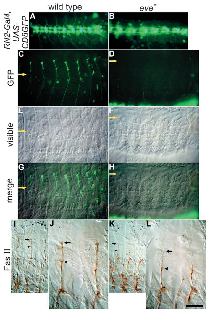
Fig. 5.
Without Eve function, most RP2 and aCC axons do not reach the muscle field. The combination of RN2-Gal4 and UAS-CD8GFP transgenes (two copies each) was placed in either a wild-type (left column) or a ΔRP2A/ΔRP2C mutant background (right column). Stage 16 embryos are shown, anterior towards the left, and dorsal upwards (except A and B, which are centered on the ventral midline). (A,B) Overview of the CNS. (C,D) GFP in the muscle field. Note that in the mutant, only a few axons are visible, and that they do not reach to the dorsal muscle field. The yellow arrow indicates the same lateral position in all panels (D,F,H are a more ventral view in order to show the small amount of axonal outgrowth that occurs near the edge of the CNS). (E,F) Nomarski view of C,D, respectively. (G) Merged image of C and E. (H) Merged image of D and F. (I–L) Anti-Fas2 staining. (J,L) Higher magnification of I,K, respectively. Note the attachment of some axons to DO2 muscles in both the wild type and the mutant (arrowheads; neuromuscular junctions to DA2 are also present, but are not visible here), but that attachments to DO1 and DA1 (only DO1 is visible here) are barely formed in the mutant (arrows). Scale bars in B and L (equal in size): 50 µm in A–I,K; 20 µm in J,L.

