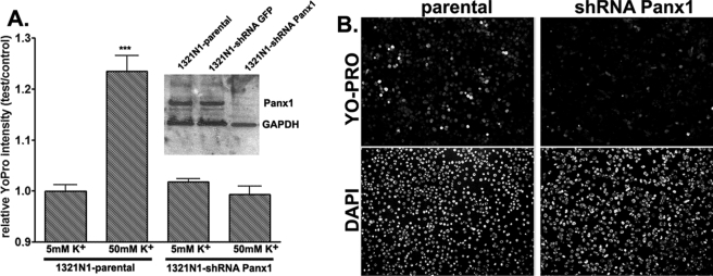
FIGURE 3.
Pannexin 1-mediated membrane permeabilization. A, relative YoPro fluorescence intensity (test/control, mean ± S.E., n = 6, two fields from three independent experiments) measured from parental and shRNA-Panx1 1321N1 cells treated for 30 min at 37 °C with normal (5 mm) and high (50 mm) K+ solutions (osmolalities adjusted by reducing equimolar amounts of NaCl). After treatments, cells were fixed and counterstained with 4′,6-diamidino-2-phenylindole. Fluorescence intensity was measured from the whole field of view (10× objective) and normalized to that obtained under control (5 mm K+) conditions. Inset, Western blot showing expression of Panx1 in parental 1321N1 cells and in two stable clones, one expressing an irrelevant shRNA (shRNA-green fluorescent protein (GFP)) and another shRNA-Panx1. B, representative images of YoPro- and 4′,6-diamidino-2-phenylindole (DAPI)-stained parental and shRNA-Panx1 1321N1 cells exposed to 50 mm K+ solution. GAPDH, glyceraldehyde-3-phosphate dehydrogenase.

