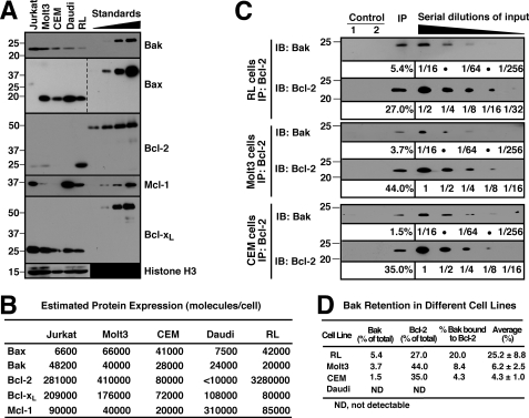
FIGURE 6.
Bak binding to Bcl-2 varies among lymphoid cell lines. A, whole cell lysates from 3 × 10⁵ of Jurkat, Molt3, CEM, Daudi, and RL cells were probed for Bak, Bax, Bcl-2, Mcl-1, and Bcl-xL. Purified GST-BakΔTM (0.1, 0.2, 0.4, and 0.8 ng), GST-BaxΔTM (0.3, 0.6, 1.2, and 2.4 ng), GST-Bcl-2ΔTM (15, 30, 60, and 120 ng), S peptide-tagged Mcl-1ΔTM (0.25, 0.5, 1.0, and 2.0 ng), and GST-Bcl-xLΔTM (0.6, 1.2, 2.4, and 4.8 ng) were used for immunoblot quantitation. Histone H3 was used as loading control. Additional blots of whole cell lysates are presented in supplemental Fig. S7. B, from the experiments shown in A and supplemental Fig. S7, the Bcl-2 family (Bak, Bax, Bcl-2, Mcl-1, and Bcl-xL) numbers per cell were calculated in Jurkat, Molt3, CEM, Daudi, and RL cells. C, CHAPS lysates from RL, Molt3, and CEM cells were immunoprecipitated (IP) with anti-Bcl-2 antibody and compared with serial dilutions of the input. Two different controls were used at the same time: beads + lysate without antibody (Control 1) and no lysate (Control 2). IB, immunoblot. D, from the experiments shown in C, the percentage of Bak bound by Bcl-2 was calculated in RL, Molt3, and CEM cells. Mean ± S.D. from three independent experiments is also shown. For Daudi, Bcl-2 is not detectable (N.D.) in either whole cell lysates (supplemental Fig. S7A) or the immunoprecipitates (data not shown).

