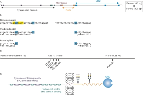
FIGURE 1.
Prolectin gene structure and protein organization. A, scale diagram of the prolectin gene. For clarity, exons are shown expanded 3-fold relative to introns. B, details of unusual GC splice site at the 5′ end of the final intron. The predicted GT splice site in the Celera annotation of the genome, GenBankTM accession EAW84437, leads to an insertion of 12 bases corresponding to insertion of 4 amino acids in the predicted protein sequence, but it leaves the reading frame intact so that the conserved residues of the CRD are present. C, scale diagram showing the relative positions of glycan-binding receptors containing C-type CRDs encoded on human chromosome 19. D, distribution of functional domains in prolectin. Predicted N-glycosylation sites are indicated with hypothetical glycans in stick figures.

