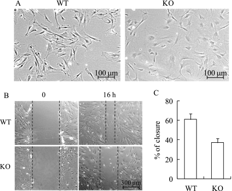
FIGURE 5.
MN1 knock-out osteoblasts have altered morphology and decreased motility. A, representative phase contrast images of sub-confluent WT and MN1 KO primary osteoblasts. B, MN1 wild-type or knock-out cells were grown to confluence, and linear scratches were created. Pictures were taken 0 and 16 h after injury. The black dashed lines mark the wound edges. C, the percentage of wound closure was quantified and presented as the mean ± S.D. (n = 9 in each group).

