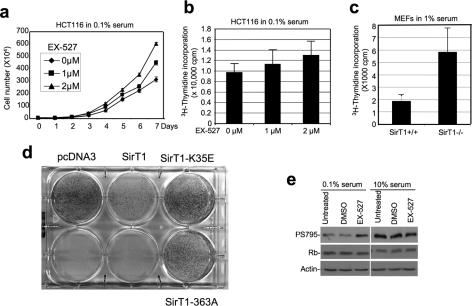
FIGURE 2.
Inhibition of SirT1 stimulates cell proliferation. a, HCT116 cells cultured in 0.1% serum were maintained in different concentrations of SirT1-specific inhibitor EX-527, and cell number was determined at the indicated time points. Error bars represent mean ± S.D. (n = 4). b, HCT116 cells cultured in 0.1% serum were treated with EX-527 for 18 h. DNA replication was measured by [3H]thymidine incorporation assay. This is a single experiment representative of three replicates. Error bars represent mean ± S.D. (n = 14) and p = 0.03 as calculated with Student's t test and p < 0.05 is statistically significant. This suggests an increase in proliferation by EX-527. c, early passage (P4) MEFs from SirT1−/− and SirT1+/+ mouse embryos were also tested for DNA synthesis rate by [3H]thymidine incorporation. An identical number of cells from each genotype was plated prior to 18-h culture in 1% serum and 1 h metabolic labeling. Error bars represent mean ± S.D. (n = 6, p = 0.00001). d, HCT116 cells were transfected with pcDNA3 vector, SirT1, SirT1-K35E NLS mutant, or SirT1-H363A deacetylase mutant. The cells were subjected to selection by G418, and the colonies were stained with Crystal Violet after 2 weeks. e, HCT116 cells treated with 2 μm EX-527 were analyzed for phosphorylation level of pRb at S795 by immunoprecipitation Western blot, followed by reprobing with Rb antibody.

