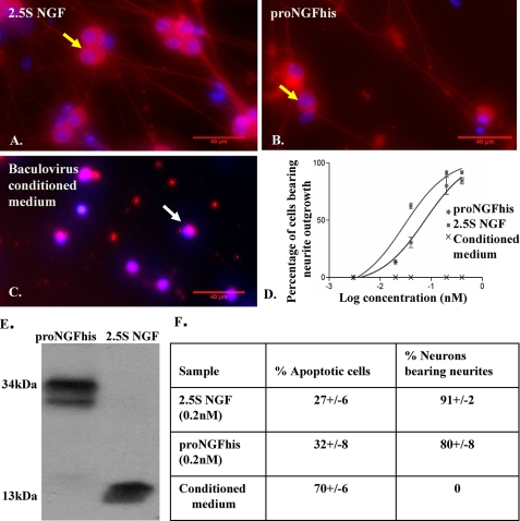
FIGURE 1.
Neurotrophic activity of proNGFhis in SCG neurons. Mouse SCG neurons were plated as described under “Experimental Procedures” and cultured in serum-free medium containing 2.5S NGF (0.4 nm) (A), proNGFhis (0.4 nm) (B), or baculovirus-infected insect cell-conditioned medium (C) for 36 h. Fixed cells were stained with TUJ-1 antibody (red) and Hoechst (blue). The scale bar is 40 μm. Yellow arrows show representative live cells and white arrows show representative dead cells. D, dose-response curve for neurite outgrowth assay. R2 = 0.96 for both proNGFhis and 2.5S NGF. Error bars represent mean ± S.E. E, Western blotting of conditioned medium using NGF antibody (MC51). There was less than 2% cleavage of proNGFhis after the neurite outgrowth assay. F, there was no significant difference between proNGFhis and 2.5S NGF in inducing apoptosis at 0.2 nm concentration (p > 0.05, Student's t test). Results were obtained from three independent experiments.

