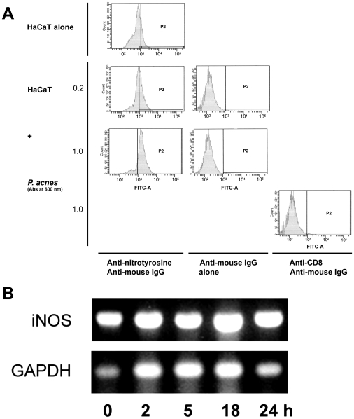
Figure 6. Nitrosyl residue formation and iNOS expression in P. acnes-stimulated keratinocytes.
HaCaT cells were incubated for 18 h with P. acnes (A600 nm = 0.2 and 1.0) and harvested following trypsine treatment after removal of bacteria. (A) Cells were incubated with primary mouse monoclonal antibody to nitrotyrosine or with anti-CD8 monoclonal antibody as control. Bound antibodies were detected using a goat anti-mouse IgG-FITC secondary antibody. Cells were then analyzed by flow cytometry as described in Materials and Methods. (B) Total RNA was extracted and the iNOS and GAPDH mRNA expression was analysed by RT-PCR. PCR fragments were visualized under U.V. on a 1.7% agarose gel after staining with ethidium bromide (1 µg/ml).

