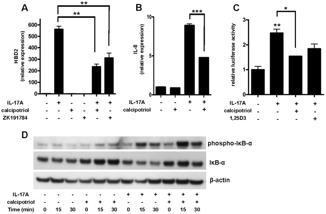
Figure 2. Induction of HBD2 and IL-8 by IL-17A is decreased by calcipotriol through inhibition of NF-κB signaling.
(A) NHEK were stimulated with IL-17A (10 ng/ml) in the presence or absence of vitamin D analogs calcipotriol or ZK191784 (10−8 M). Cells were harvested after 24 h and HBD2 transcript levels were analyzed by qPCR. In (B) IL-8 transcript expression after stimulation of NHEK with IL-17A (10 ng/ml) in the presence or absence of calcipotriol (10−8 M) is displayed. Data are means±SD of a single experiment performed in triplicate and representative of 2 to 3 independent experiments (**P<0.01, ***P<0.001; Student's t test). (C) To study involvement of the Nf-κB pathway we performed reporter gene analyses with an Nf-κB reporter plasmid. 24 h after transfection of HaCaT keratinocytes cells were stimulated with IL-17A (10 ng/ml) in the presence or absence of calcipotriol or 1,25D3 (10−8 M). Luciferase activity was assayed (*P<0.05, **P<0.01; Student's t test). (D) To further confirm Nf-κB involvement cells were stimulated with IL-17A (10 ng/ml) in the presence or absence of calcipotriol (10−8 M) and harvested after 0, 15 or 30 min. Western blot analysis using antibodies against phospho-IκB-α and unphosphorylated IκB-α were performed to analyze activation of NF-κB. Staining for β-actin served as a loading control.

