
Figure 4. Cortactin promotes tumor vascularization.
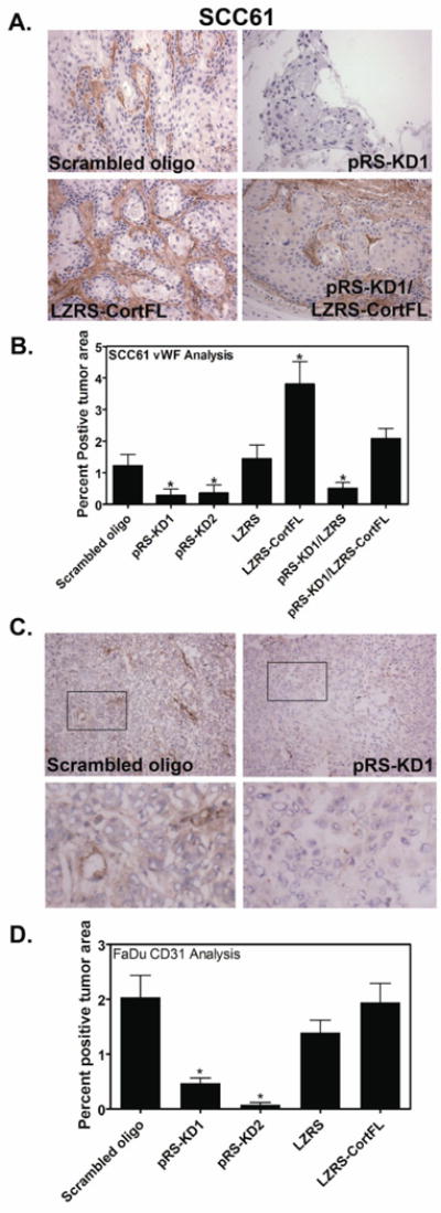
Tumor vascularization was assessed by staining with an antibody against for von Willebrand Factor (vWF), a coagulation factor that underlies endothelial cells in large vessels or with an antibody recognizing the endothelial cell marker CD31 to identify small vessels. Cortactin expression strongly correlates with the presence of large vessels in SCC61 cells (A and B) and small vessels in FaDu cells (C and D). A. Representative images of SCC61-derived tumors stained for von Willebrand Factor (vWF). Positive staining is brown and all nuclei are counterstained light blue. 20× images are shown. Scale bar = 100 μm. B. Quantification of vWF stained tumors for all cell lines is shown as percent positive tumor area as determined by color thresholding in Metamorph. Data are represented as mean ± SEM. C. Representative images of FaDu-derived tumors stained for CD31. Positive staining is brown and all nuclei are counterstained light blue. 20× images are shown. Scale bar = 100 μm. D. Quantification of CD31-stained tumors for all cell lines is shown as percent positive tumor area as determined by color thresholding in Metamorph. Data are represented as mean ± SEM. Asterisks indicate p<0.05 compared to scrambled cortactin oligo control.