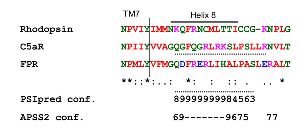
Figure 9. Computational prediction of the helical structure in the membrane proximal region of the cytoplasmic tail of FPR.
Amino acid sequences of bovine rhodopsin (N301-G328), human C5aR (N296-T324) and human FPR (N297-T325) were aligned (vertical line) relative to the 7th transmembrane domain (TM7). The position of helix 8 of rhodopsin is based on the resolved crystal structure [17]. Star (*) indicates identical amino acids in all three sequences. Two dots (:) indicates tolerable amino acid substitutions, and one dot (.) indicates amino acid residues of similar size. The numbers underlying the sequences represent the rate of confidence for helical structure provided by PSIpred and APSS2 servers. Higher number stands for higher confidence. Hyphen (-) represents the highest confidence of 10, used in the APSS2 prediction. Dotted line (…..) underlines the sequence of the putative helix 8, as predicted by PSIpred and APSS2 analysis.

