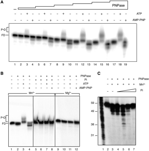
Figure 3.
Binding and degradation of ssDNA by PNPase. In (A) [γ32P]-ssDNA60 (1 nM) was incubated with increasing concentrations of PNPase (0.05, 0.1, 0.25, 0.5, 1.5 and 2 nM) for 30 min in buffer B containing 1 mM ATP, AMP-PNP or lacking a nucleotide cofactor, and the samples were separated in 10% nPAGE. FD, free DNA; PD, PNPase–DNA complexes. In (B) [γ32P]-ssDNA60 (1 nM) was incubated with PNPase (2 nM) for 30 min in buffer B (Mn2+) or buffer C (Mg2+) containing 1 mM ATP, AMP-PNP or lacking a nucleotide cofactor, and with or without added 2 mM Pi, and the samples were separated in 10% nPAGE. In (C) [γ32P]-ssDNA60 (1 nM) was incubated with PNPase (0.3 nM) for 30 min in buffer B containing 1 mM AMP-PNP and no added Pi, or increasing Pi (0.2, 2, 20 and 200 μM), and the samples were separated in 15% dPAGE.

