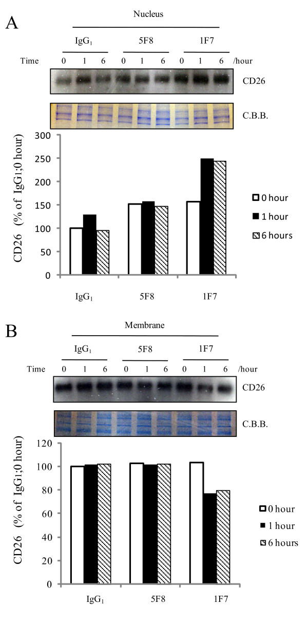
Figure 3.
Increase of CD26 in nucleus following treatment with CD26 mAb, 1F7. J/CD26 cells were incubated with media containing the CD26 mAbs, 1F7 or 5F8, or control mouse IgG1. After incubation for 0, 1, or 6 hours, the cells were separated into (A) nuclear and (B) membrane fractions and CD26 proteins were immunoblotted. Coomassie brilliant blue (C.B.B.) staining was shown. Representative CD26 expression was shown. Similar results were obtained in three independent experiments.

