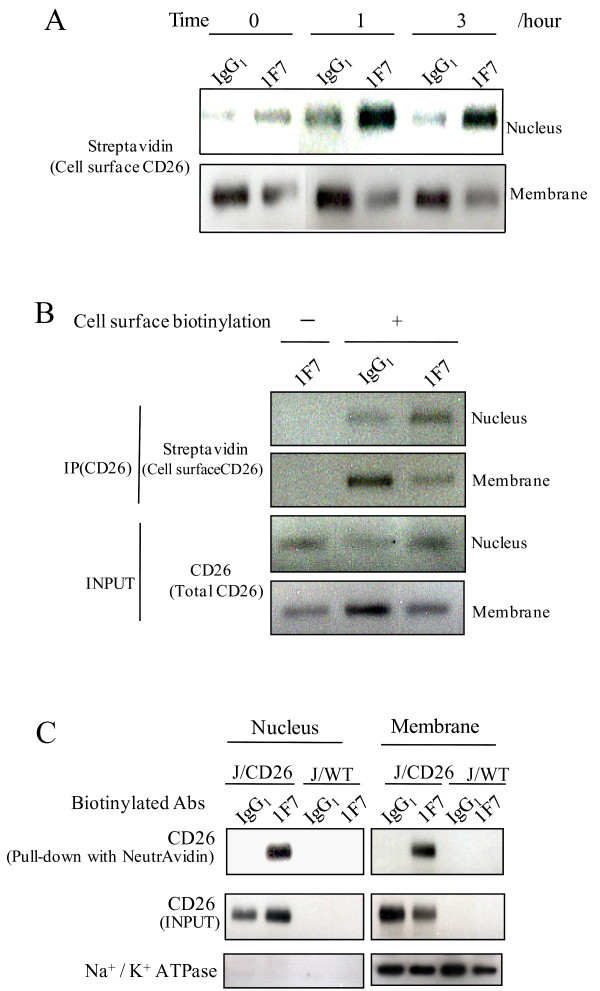
Figure 4.
Translocation of CD26 into nucleus from cell surface. (A and B) Cell surface biotin-labeling of (A) J/CD26 cells and (B) Karpas 299 cells. Cells were treated with/without sulfo-NHS-biotin, and incubated with 1F7 or IgG1 for (A) 0, 1 and 3 hours, or for (B) 1 hour. The cells were separated into nuclear and membrane fractions, immunoprecipitated with CD26 mAb, and immunoblotted. (C) J/Wt cells and J/CD26 cells were treated with biotin-labeled antibodies (1F7 or control IgG1) and separated into nuclear and membrane fractions. The biotinylated antibody-bound proteins were purified on beads immobilized to NeutrAvidin and separated by SDS-PAGE. Representative CD26 expression was shown. Similar results were obtained in three independent experiments.

