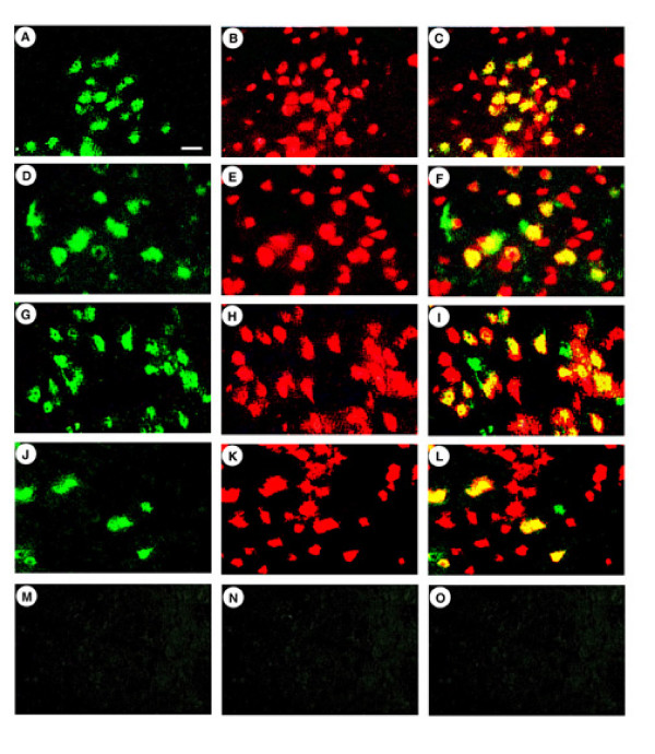
Figure 3.
β-gal-IR positive striatal cells that also contain NeuN-IR from rats sacrificed at 4 days or 2 months after gene transfer with pNFHlac packaged using specific combinations of mutated HSV-1 proteins. β-gal-IR was detected using a rabbit anti-E. coli β-gal antibody, and was visualized using a fluorescein isothiocyanate-conjugated goat anti-rabbit IgG. In the same sections, NeuN-IR was detected using a mouse monoclonal anti-NeuN, and was visualized using a rhodamine isothiocyanate-conjugated goat anti-mouse IgG. NeuN is a neuronal marker found in the nucleus. A-C. pNFHlac/Δul46&47, ul13g, rat sacrificed at 4 days; β-gal-IR (A), NeuN-IR (B), merged (C). Many of the β-gal-IR cells also contain NeuN-IR. D-F. pNFHlac/Δul46&47, ul13g, rat sacrificed at 2 months; β-gal-IR (D), NeuN-IR (E), merged (F). G-I. pNFHlac/VP16in14, gene12, ul13g, rat sacrificed at 4 days; β-gal-IR (G), NeuN-IR (H), merged (I). J-L. pNFHlac/VP16in14, gene12, ul13g, rat sacrificed at 2 months; β-gal-IR (J), NeuN-IR (K), merged (L). M-O. pNFHlac/VP16in14, gene12, ul13g, rat sacrificed at 4 days; no primary antibodies. Scale bar: 25 μm.

