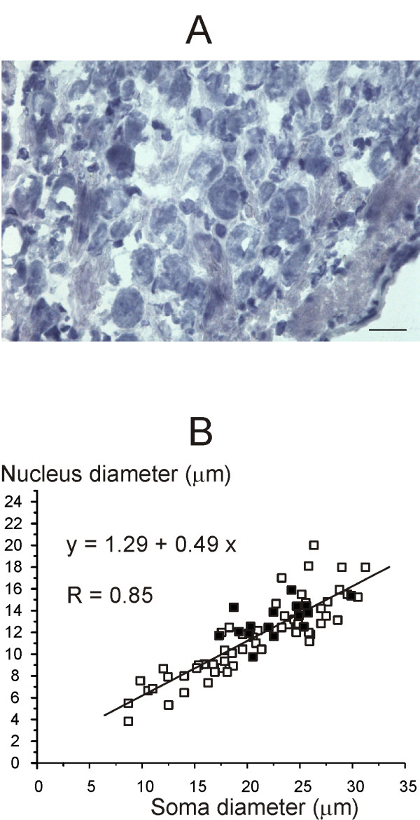
Figure 5.

Scarpa's ganglion neurons size. A: Photomicrograph of a typical ganglion slice counterstained with ematoxylin/eosin, which stains both nucleus and cytoplasm. Calibration bar: 20 μm. B: scatter plot showing neuronal soma and relative nucleus diameter in a typical slices stained with hematoxylin/eosin (squares) and in a typical slice stained for the H3R antibody (filled squares). The equation for the regression line and the correlation coefficient value are shown in figure.
