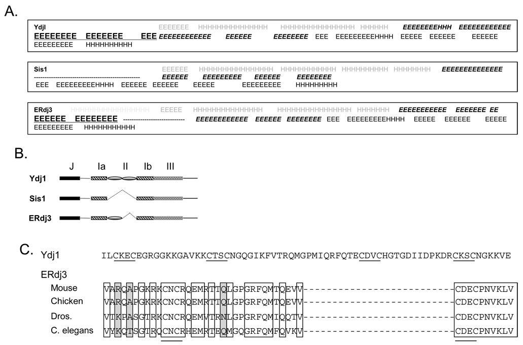
Figure 1. Comparison of ERdj3 with Ydj1 and Sis1.
(A) Secondary structure predictions for ERdj3, Sis1 and Ydj1. Colors represent the different domains: Light grey: ER targeting sequence, grey: domain J, bold and italics: domain I, bold and underlined: domain II, black: domain III, H: α helix, and E: β sheet. (B) The schematic representation domain structures of Ydj1, Sis1 and ERdj3 showing that Sis1does not contain a cysteine-rich domain II and that ERdj3 is more similar to Ydj1, although it contains an atypical, smaller domain II. (C) Domain II sequences of ERdj3 from various species were aligned and compared to domain II of Ydj1. Clear boxes indicate amino acid identities, whereas grey shading indicates amino acid similarity. Cysteine residues (CysXXCys motif) are underlined.

