Figure 8.
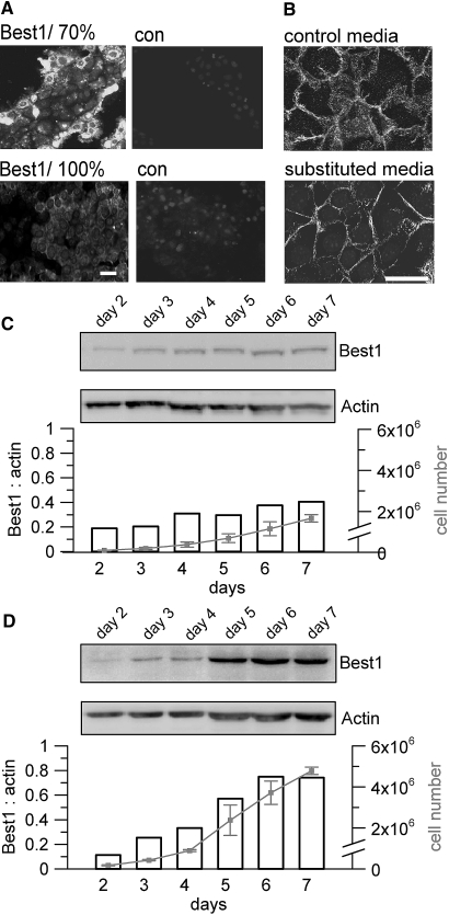
Cell density controls expression of Best1. (A) Immunohistochemistry of Best1 in mouse M1 cells. When grown to 70% density, cells at the edge of an island show high levels of Best1 expression, while cells in the center of islands and monolayers grown to 100% confluence downregulate Best1 expression (n = 5 for each series). (B) β-catenin localization in cells grown in normal media (control media) or cells exposed to conditioned medium from confluent monolayers (substituted media) (n = 3 for each series). Membrane-limited expression of β-catenin suggests MET in cells exposed to conditioned medium (scale bars = 20 μm). (C) Reduced cell proliferation and Best1-expression in cells grown in media substituted by supernatant (50 vol%) from confluent M1 cultures. (D) Enhanced proliferation and Best1-expression in cells grown in media that was frequently (twice per day) replaced (n = 2 for each series).

