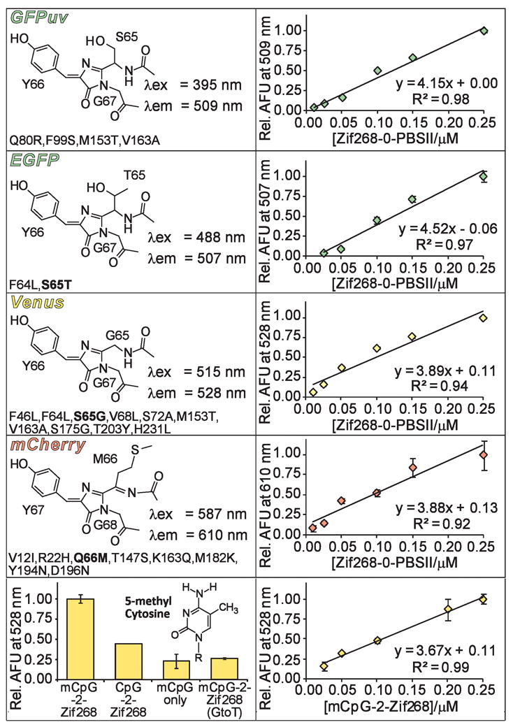
Figure 2.

Properties of FPs utilized in SEER. The left panels display chromophore structures and wavelengths of maximum excitation (λex) and emission (λem) for each of the full length FP variants. Mutations (A. victoria numbering) are listed below the corresponding structures with chromophore mutations indicated in bold. mCherry includes additional GFP-type residues on both its N- and C-terminus.19 The right panels show a DNA titration for each SEER-FP system. The bottom panels show specificity data and a DNA titration for the mCpG-SEER system.
