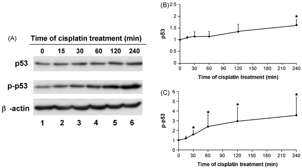
Fig. 1.
Early p53 activation during cisplatin treatment. RPTC cells were incubated with 20 µM cisplatin for 0–240 min. Whole cell lysate was collected for immunoblot analysis of total p53 and phosphorylated p53 (p-p53). The blots were then reprobed for β-actin to monitor protein loading and transferring. (A) Representative immunoblots. (B) Densitometry of p53. (C) Densitometry of p-p53. For densitometric analysis, the p53 or p-p53 signals of control cells was arbitrarily set as 1 in each blot, and the signals of experimental conditions in the same blot were normalized with the control to show their p53 or p-p53 levels. The results were from at least three immunoblots of separate experiments. Data are expressed as mean ± S.D. (n ≥ 3). *Statistically significantly different from the control.

