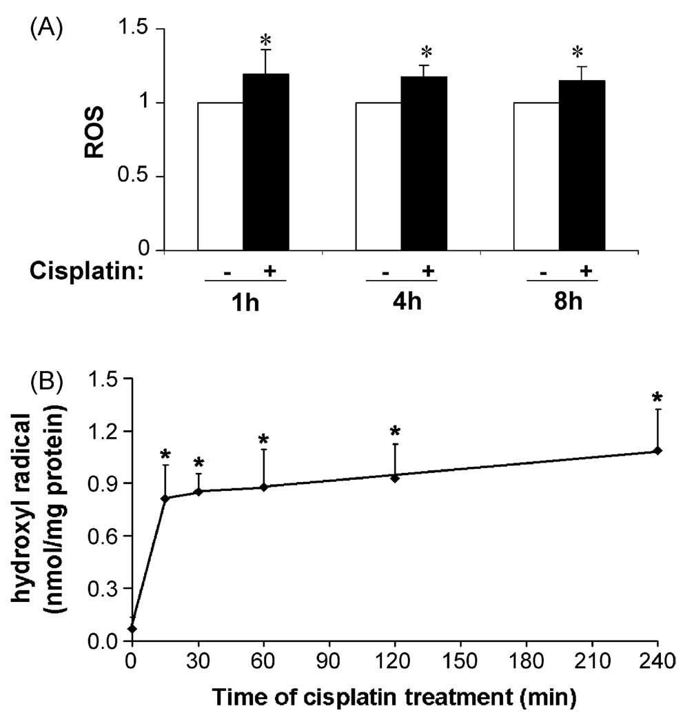
Fig. 2.
ROS and hydroxyl radical accumulation during cisplatin treatment. RPTC cells were incubated with 20 µM cisplatin for indicated time. (A) ROS accumulation. ROS was measured using the fluoregenic dye DCF as described in Section 2. The fluorescence signals of the experimental samples were normalized with the values of the control, which was arbitrarily set as 1. (B) Hydroxyl radical accumulation. Hydroxyl radicals were measured by the deoxyribose degradation assay as detailed in Section 2. Data are expressed as mean ± S.D. (n = 4). *Statistically significantly different from the control group without cisplatin treatment. The results demonstrate a marginal increase of total ROS and a drastic accumulation of hydroxyl radicals during cisplatin treatment.

