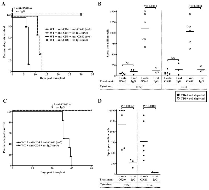
Figure 2. Stimulatory anti-OX40 mAb acts on CD4+ T cells.
(A) WT C57BL/6 allograft recipients were transiently depleted of CD4+ or CD8+ cells (1 mg i.p. anti-CD4 mAb or anti-CD8 mAb days -1, 0, and 7 relative to transplant) concurrent with agonistic anti-OX40 mAb (closed symbols, solid line) or control rat IgG treatment (open symbols, dashed line) (0.25 mg i.p. days 0, 1, 3 and 7 relative to transplant). (B) Recipients treated as described in Panel A were sacrificed either at time of rejection or 30 days post transplant. Donor-reactive Th1 (IFNγ) and Th2 (IL-4) splenocyte responses were quantified by ELISPOT. Individual data points represent the number of primed donor-reactive Th1 or Th2 cells in individual allograft recipients. Bars are indicative of the average number of donor-reactive splenocytes per experimental group. (C) WT recipients inductively depleted of CD4+ T cells were treated with stimulatory anti-OX40 mAb (closed circles, solid line) or control rat IgG (open circles, dashed line) 30 days post transplantation (0.25 mg i.p. days 30, 31, 33 and 40 relative to transplant) and monitored for function. (D) Recipients treated as described in Panel C were sacrificed either at the time of allograft rejection or 30 days after the initiation of treatment. Th1 (IFNγ) and Th2 (IL-4) splenocyte responses were quantified by ELISPOT. Individual data points represent the donor-reactive Th1 and Th2 responses of individual allograft recipients. Bars are indicative of the average number of donor-reactive Th1 or Th2 cells per experimental group.

