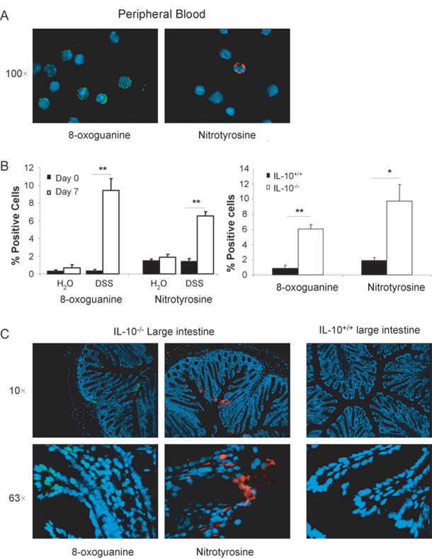
Fig. 6. 8-oxoguanine and Nitrotyrosine Formation in Peripheral Leukocytes and the Colon.
A. Representative images of positive staining for 8-oxoguanine (green, left) and nitrotyrosine (red, right) in leukocytes of DSS-treated wildtype (7 days) and IL-10−/−mice (6 months). B. Percent positive cells for 8-oxoguanine and nitrotyrosine staining before and after DSS treated mice (7 days), n=6 per group (LEFT) and in IL-10−/− mice (6 months), n=4 per group (RIGHT). *: p<0.05, **: p<0.01 by Student’s unpaired t-test. C. Representative images of 8-oxoguanine (green) and nitrotyrosine (red) in colon sections of IL-10−/− mice (6 months) and wildtype mice.

