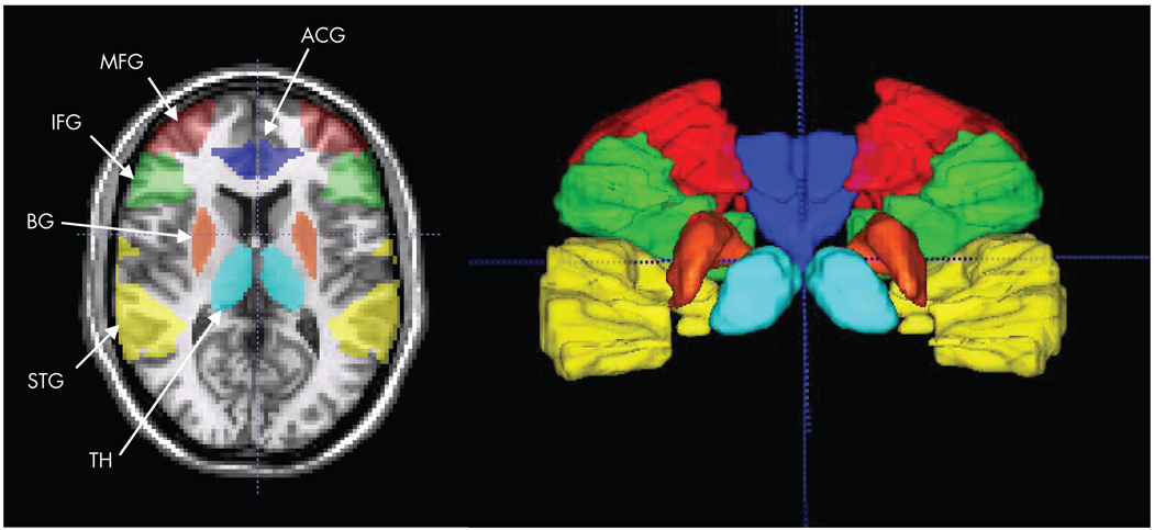
FIGURE 1. An Axial Slice and a 3-D Rendering (Posterior View; Cross Hair at AC) Show the Six Regions of Interest Selected Based on a priori Hypotheses of Automatic and Controlled Auditory Processing.
Regions are color coded: dark blue=anterior cingulate gyrus (ATG), green=inferior frontal gyrus (IFG), red=middle frontal gyrus (middle frontal gyrus), yellow = superior temporal gyrus (STG), orange=basal ganglia (BG), and light blue=thalamus (TH).

