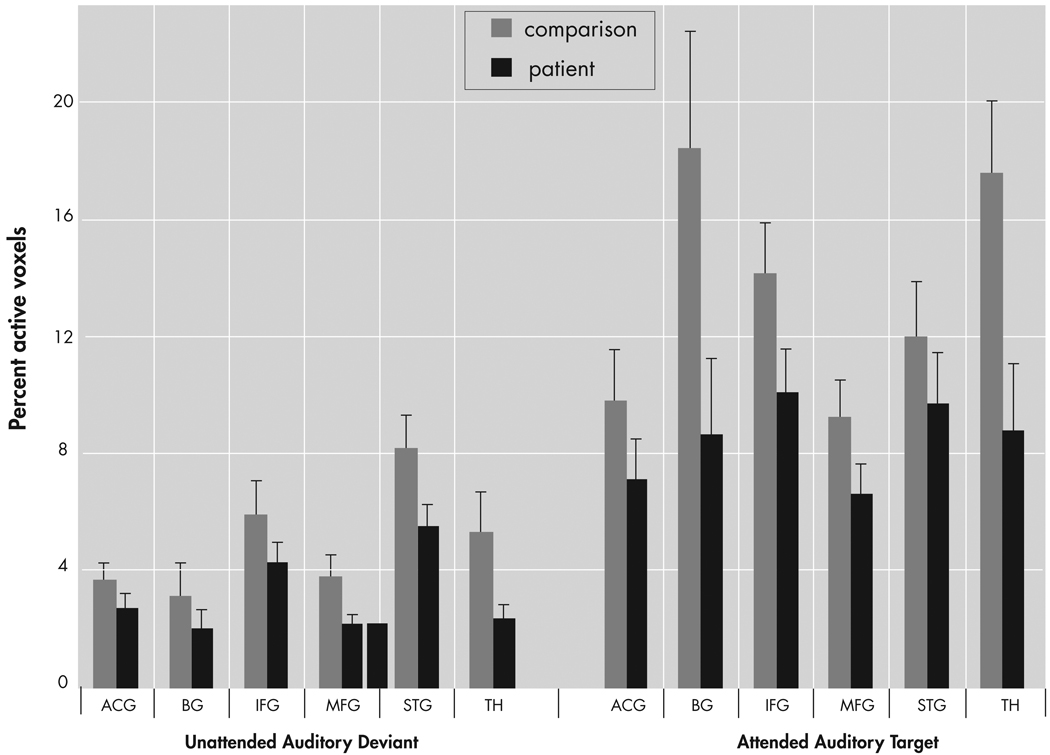
FIGURE 4. Quantitative Results of Region of Interest Analysis for Percent Active Voxels for Patients and Comparison Subjects in Six Hypothesized Regions of Interest.
Patients showed lower overall extent of activation than comparison subjects (main effect of group). Activation for the UAD condition was lower than for the AAT condition (main effect of condition). ACG=anterior cingulate gyrus; IFG=inferior frontal gyrus; MFG=middle frontal gyrus; STG=superior temporal gyrus; BG=basal ganglia; TH=thalamus.

