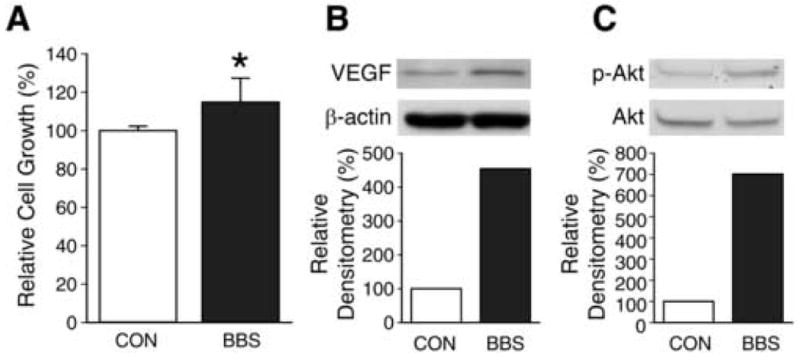
Figure 1. BBS stimulates cell growth, VEGF and p-Akt expression in SK-N-SH cells.

(A) Effects of BBS (10-7 M) on SK-N-SH cell viability were determined using CCK-8 kit (Data represent mean ± SEM values of eight replicate experiments; * p < 0.05 vs. control). (B) SK-N-SH cells were treated with BBS (10-7 M) for one day after overnight serum-free conditions. Cell lysates were prepared and analyzed by Western blot for VEGF. (C) SK-N-SH cells were treated with BBS (10-7 M) for 5 min after overnight serum-free condition. Cell lysates were prepared and analyzed by Western blot for p-Akt and total Akt. Experiments were repeated on two separate occasions.
