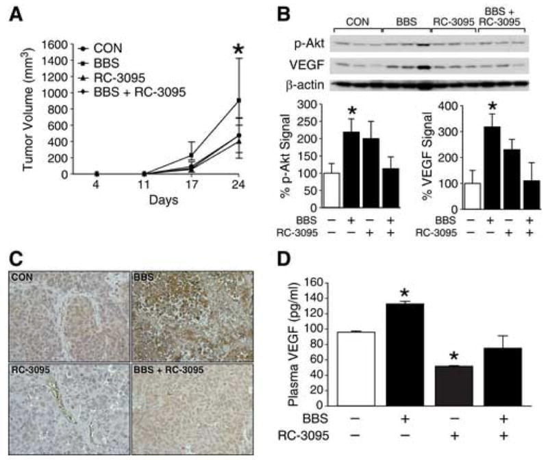
Figure 4. GRP antagonist inhibits BE(2)-C tumor growth and angiogenesis.

(A) Tumor volumes in nude mice with BE(2)-C cells treated with vehicle, BBS (20 μg/kg/injection; s.c., t.i.d.), and/or RC-3095 (10 μg/kg/injection; s.c., q 12 h), as described in “Materials and Methods” (5-6 mice/group). (B) Expression of p-Akt and VEGF in BE(2)-C tumor tissue samples (three representative tumor samples from each group are shown). (C) Representative sections of BE(2)-C tumors stained with anti-human VEGF antibody (brown); magnification, X400. (D) VEGF plasma levels from mice detected by ELISA. Data from all figures represent mean ± SEM; * p < 0.05 vs. control.
