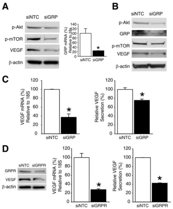
Figure 5. GRP/GRPR siRNA attenuates angiogenesis in human neuroblastoma cells.

(A) Western blot analysis of p-Akt, p-mTOR and VEGF protein expression in SK-N-SH cells at 3 d post-transfection with siNTC or siGRP (left panel). Quantitative RT-PCR for GRP mRNA levels in SK-N-SH cells at 2 d post-transfection with siNTC or siGRP (right panel). (B) Western blot analysis of p-Akt, GRP, p-mTOR and VEGF protein expression in BE(2)-C cells at 3 d post-transfection with siNTC or siGRP. (C) Quantitative RT-PCR analysis for VEGF mRNA level in BE(2)-C cells at 2 d post-transfection with siNTC or siGRP (left panel). VEGF levels in BE(2)-C using human VEGF ELISA kit. Cell culture supernatants were harvested at 3 d post-transfection with siNTC or siGRP (right panel). (D) BE(2)-C cells were transfected with siNTC or siGRPR for various assays. GRPR and VEGF protein expression was determined by Western blot analysis at 3 d post-transfection (left panel). VEGF mRNA levels were assessed by quantitative RT-PCR at 2 d post-transfection (middle panel) and VEGF levels in cell culture supernatants were measured by ELISA at 3 d post-transfection (right panel). Data from all figures represent mean ± SEM; * p < 0.05 vs. control.
