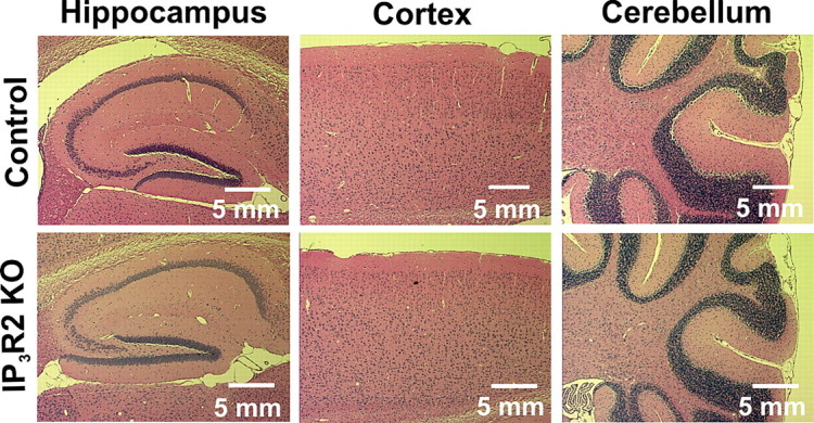
Figure 1.

Histological analysis of IP3R2 KO brains reveals no obvious abnormalities. Histological staining of brain sections were taken from littermate control (n = 3) and IP3R2 KO (n = 3) mice. Six-micrometer-thick paraffin-embedded sections were cut and stained with hematoxylin and eosin to visualize brain cytoarchitecture. No difference in the gross overall morphology or in the general cell layering was apparent between the IP3R2 KO mice and littermate controls in any brain region; data from hippocampus, cortex, and cerebellum are shown.
