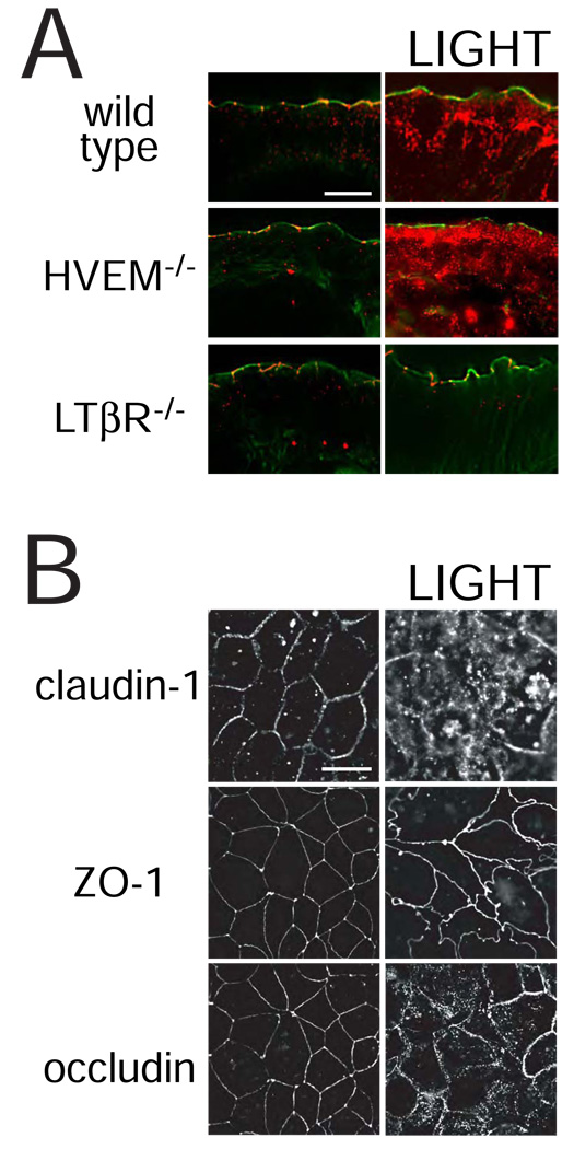
Figure 5. LIGHT-induced barrier dysfunction is accompanied by tight junction protein endocytosis.
A. Jejunal mucosa from wild type, HVEM−/−, and LTβR−/− mice were snap-frozen 3 hours after injection of LIGHT. Sections were stained for occludin (red) and F-actin (green). In control mice occludin is primarily localized to the tight junction, where it punctuates the perijunctional actomyosin ring. LIGHT induces the formation of large intracellular occludin stores in jejunal epithelia of wild type and HVEM−/− mice. In contrast, LTβR−/− mice did not internalize occludin in response to LIGHT. Results are representative of 3 independent experiments. Bar = 10 µm.
B. Caco-2 monolayers were pre-treated with IFN-γ followed by transfer to media with or without LIGHT. After 8 hours the monolayers were fixed and immunostained for claudin-1, ZO-1, and occludin. Obvious claudin-1 and occludin internalization is induced by LIGHT. Vesicular ZO-1 deposits are not formed, but the distribution at the tight junction is markedly disrupted. Results are representative of 5 independent experiments. Bar = 20 µm.

