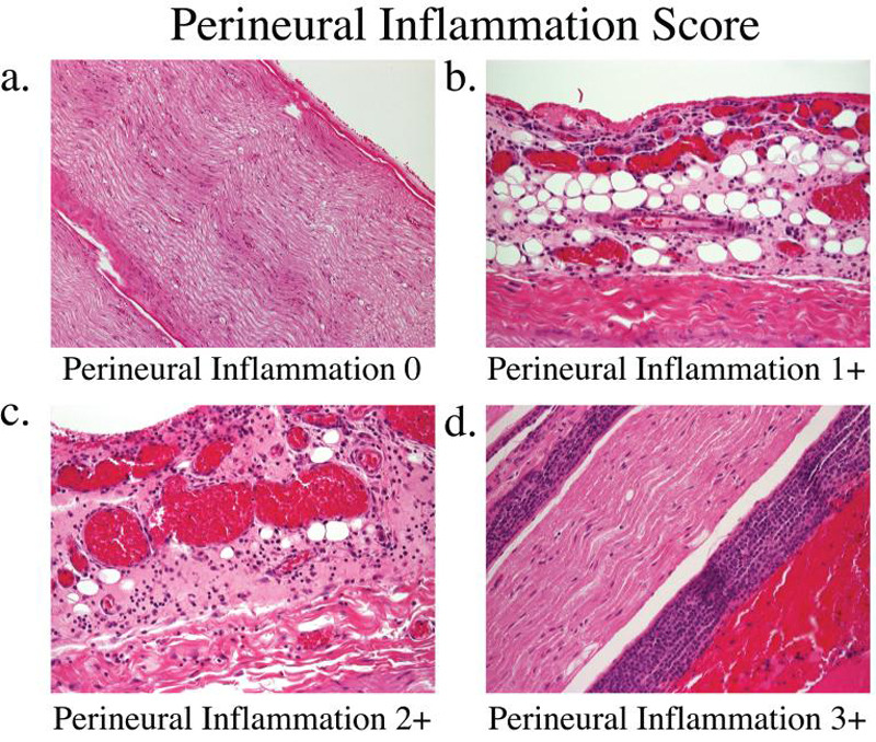
Figure 5.

Nerves were sectioned and stained with hemotoxylin and eosin to assess perineural inflammation at 24 hours and 14 days. Nerves in the bupivacaine group had higher inflammation scores at 24 hours when compared with the saline control. Bupivacaine plus dexmedetomidine and dexmedetomidine alone had similar inflammation scores compared with normal saline at 24 hours. At 14 days, nerves in both groups were completely normal with inflammation scores of 0. (A) Inflammation score = 0: The perineural space is void of any significant inflammatory cells. (B) Inflammation score = 1: Focal portions of perineural inflammation involving 5-10% of the sections. (C) Inflammation score = 2: Moderate degree of perineural inflammation. (D) Inflammation score = 3: Severe inflammation is seen with large numbers of lymphocytes surrounding the nerve.
