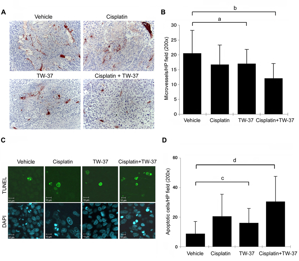
Figure 6.
Effect of cisplatin and/or TW-37 on tumor angiogenesis and apoptosis. Each SCID mouse was implanted with two scaffolds seeded with 1.0 × 10⁵ OSCC-3 and 9.0 × 10⁵ HDMEC. Eighteen days after implantation, mice were randomly assigned to four groups (n=14 tumors per experimental group) as follows: 5 mg/kg cisplatin on day 0 and day 5 via intraperitoneal injection; 15 mg/kg TW-37 daily for 5 days via intraperitoneal injection; combination of the regimens above for cisplatin and TW-37 for 5 days; or vehicle injected controls. Mice were euthanized on day 6, i.e. one day after the end of treatment. A,B, Tissue sections were stained for Factor VIII (red color) and counterstained with hematoxylin. Factor VIII positive vessels were counted under light microscopy at 200× magnification. C,D, Tissue sections were stained with in situ TUNEL and with DAPI. Images were prepared at 200× magnification and TUNEL positive cells were counted using the Image J software. Statistical significance (P<0.05) is depicted by lowercase letters, as follows: (a) blood vessel density is significantly lower in cisplatin or TW-37 treated tumors than in control tumors; (b) blood vessel density is significantly lower in combination treatment than in any other condition. (c) number of apoptotic cells is significantly higher in cisplatin or TW-37 treated tumors than in control tumors; (d) number of apoptotic cells is significantly higher in combination treatment than in any other condition.

