
Figure 5.
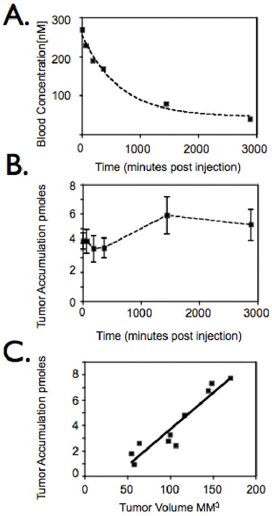
In vivo characterization of IPL-NP. A. Mice bearing tumors derived from LNCaP cells were injected with IPL-NP (20 mg/kg Fe) then blood analyzed for agent presence at indicated time points post injection. B. Time course of IPL-NP accumulation into LNCaP derived tumors. Mice bearing LNCaP derived tumors were injected with IPL-NP (20 mg/kg Fe) then imaged via FMT at 0,1,3,6,24, and 48 hours post injection. C. Mice bearing tumors of different volumes derived from LNCaP cells were injected with identical doses of IPL-NP (20 mg/kg Fe) then imaged via FMT.