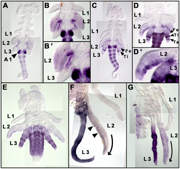
Figure 3. Expression patterns of Ubx and Abdominal-A (UbdA) proteins during G. buenoi embryogenesis.
(A) Early segmented embryo where a faint UbdA is first seen in the legs (arrowhead indicates the strong UbdA accumulation in abdominal segment A1; anterior to the top). (B–B′) UbdA accumulates uniformly throughout the entire limb bud of L2, but is absent from the distal parts of L3 limb buds. (C–G) Dynamics of UbdA accumulation in both L2 and L3 legs (arrowheads indicate UbdA accumulation in the segments of each leg). (C) UbdA expression now appears as a strong stripe in the tibia and a faint stripe in the femur of L3. (D) This expression is followed by another strong stripe that corresponds to the Tarsus in L3. (D′) In L2, the levels of UbdA accumulation are stronger in the posterior relative to the anterior compartment. (E–G) This pattern continues in later embryonic stages where UbdA becomes strong and uniform in the whole L3 legs (E,F), and also persists in distinct levels between the anterior and posterior compartments in L2 (arrowheads in F). In the trunk, UbdA is first excluded from the thoracic segments (A–C), then appears later in faint levels in both T3 and the posterior compartment of T2 (D–E). Note the dynamics of L2 and L3 size development in (E–G), where L3 first reaches a longer size F before the size of L2 catches up (G). This dynamic of leg growth is consistent with the typical arrangement of these two legs along the embryo axis. Ti: Tibia, Ta: tarsus and Fe: Femur. Curved arrows in (F) and (G) indicate the differential growth between the anterior and the posterior compartments of L2, which results in the curving of this leg.

