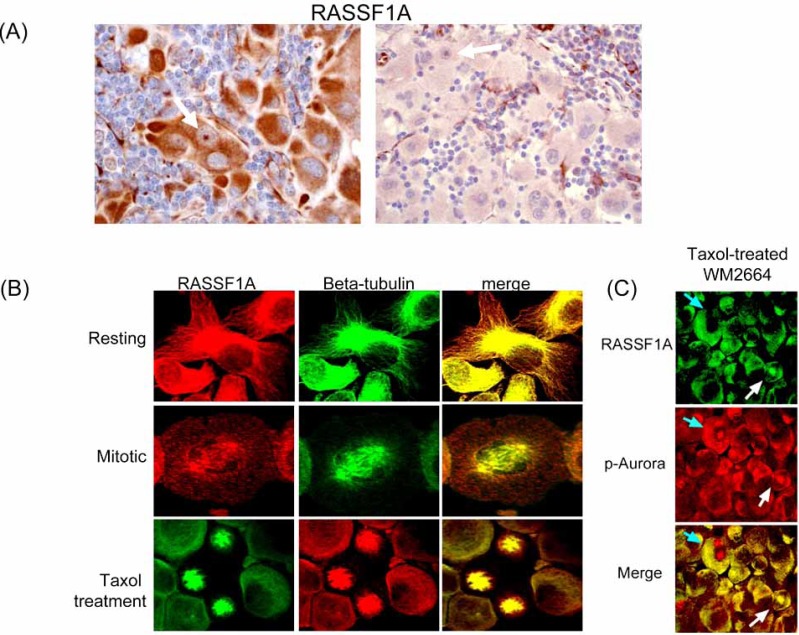
Fig. (4).
Variable RASSF1A expression in melanoma. (A) High level RASSF1A expression in the cytoplasm of a melanoma metastatic to lymph nodes is contrasted with near absence of expression in another progressed melanoma. White arrows highlight the nuclear localization of RASSF1A seen in cycling cells. Immunohistochemical staining was performed on formalin-fixed paraffin-embedded tumor sections using a mouse monoclonal antibody (eB114-10H1. eBioscience, San Diego, CA) and the ABC avidin-biotin detection method. Activated endothelium within each tissue serves as a positive control. (B) RASSF1A in non-dividing cells is present in the cytoplasm in association with the actin-tubulin cytoskeleton. During the later stages of mitosis, RASSF1A transiently colocalizes with tubulin and other spindle components at the spindle poles. Use of mitotic spindle inhibitor (paclitaxel) results in trapping of RASSF1A in stalled mitotic spindle complexes. Confocal microscopy performed with RASSF1A polyclonal antisera (N-12, Santa Cruz Biotechnology, Santa Cruz, CA), and a beta-tubulin mouse monoclonal antibody (clone DM1A, Sigma). (C) In melanoma cell lines, RASSF1A and Aurora kinases colocalize at the mitotic spindle in a subset of cells. Confocal microscopy was performed using a pan-phospho-Aurora antisera (Thr288A/Thr232B/ Thr198C, Cell Signaling Technology, Beverly, MA) and a RASSF1A mouse monoclonal antibody (eB114-10H1, eBioscience).

