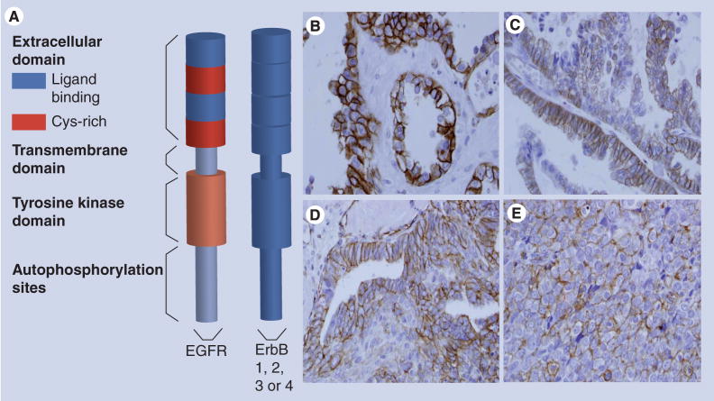
Figure 2. Activated EGF-receptor in ovarian tumors.
(A) Model of the EGFR. The extracellular N-terminal domain contains two subdomains that directly interact with ligand and two cysteine-rich subdomains. There is a single transmembrane domain that links the extracellular domain to the intracellular tyrosine kinase domain and the C-terminal tail that contains the autophosphorylation sites. The EGFR dimerizes with other ErbB receptors. (B–E) Immunohistochemical staining for activated (phospho-)-EGFR in ovarian tumors. Immunohistochemical analysis was performed retrospectively as described in [41], using antibodies to phospho-EGFR (1:400, Zymed®, CA, USA) according to standard procedures. EGFR activation (phospho-EGFR) was evident in 35% of the specimens analyzed (n = 146) and MMP-9 expression was statistically positively correlated with EGFR activation [41]. (B) Ovarian clear cell carcinoma; (C) mucinous ovarian carcinoma; (D) endometrioid ovarian carcinoma; (E) serous ovarian carcinoma.
EGFR: EGF-receptor.

