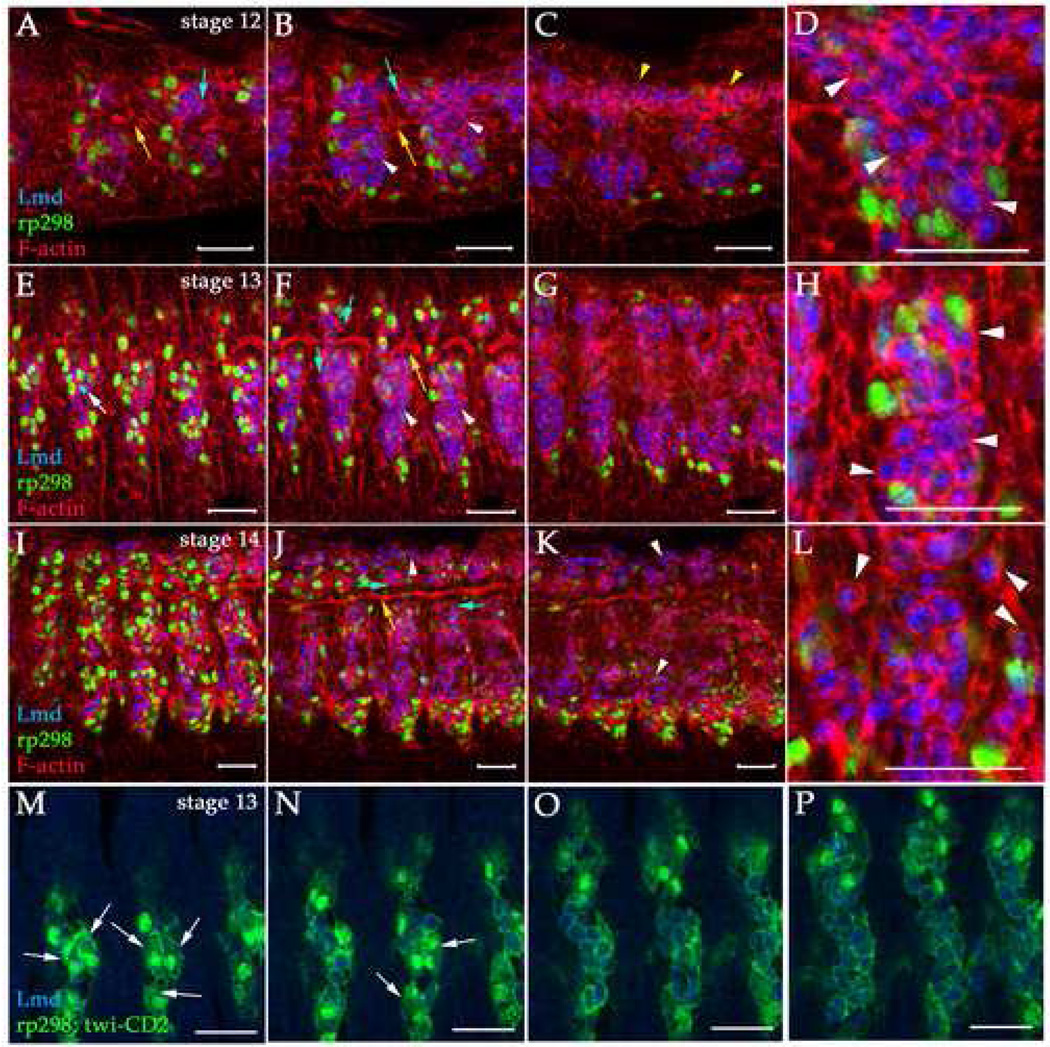
Figure 1. FC and FCM arrangements during myoblast fusion.
Single optical slices of rp298-lacZ (A–L) or rp298-lacZ; twi-CD2 (M–P) stage 12 (A–D), 13 (E–F, M–P) and 14 (I–L) embryos labeled with anti-βgal to label FC/myotube nuclei (green), anti-Lmd to label FCMs (blue), phalloidin to label F-actin (red) and anti-CD2 to label mesodermal cell membranes (green) are shown. Panels on the left (A, E, I, M) are more external than those on the right (C, G, K, P). Panels D, H and L show close-ups of FCMs in panels B, F and J respectively. Dorsal is up and anterior is left and scale bars are 20 µm in all panels. The developing trachea (yellow arrows, A, B, F, H, K) were used for accurate staging of embryos (Manning and Krasnow, 1993). Mesodermal hemisegments narrow along the A–P axis and extend along the D–V axis during these stages due to germband retraction and dorsal closure (compare A–C with I–K). FCs/myotubes (green) are present in more external panels (A, E, I, M, N), but not more internal where the majority of FCMs (blue) are located (B, C, F, G, J, K, O, P). (A–H) FCs and FCMs are tightly packed together at stages 12 and 13 (white arrowheads, B–D, F–H, M–P). (I–L) However, during stage 14 the FCMs separate from one another and round up to become migratory (white arrowheads, J–L). FCMs in similar locations contact different cell types such as other FCMs, FCs/myotubes and epithelial cells (blue arrows, A, B, F, J). Fusion events can be visualized by colocalization of rp298-lacZ and Lmd (white arrow, E) and can be visualized using this approach from stage 13 onwards. (M–P) Myotubes containing two nuclei can be observed in external cell layers at late stage 13. No FCMs are observed in these cells layers at this stage. The visceral mesoderm is beneath the somatic mesoderm at stage 12 and expresses high levels of F-actin (yellow arrowheads, C).

