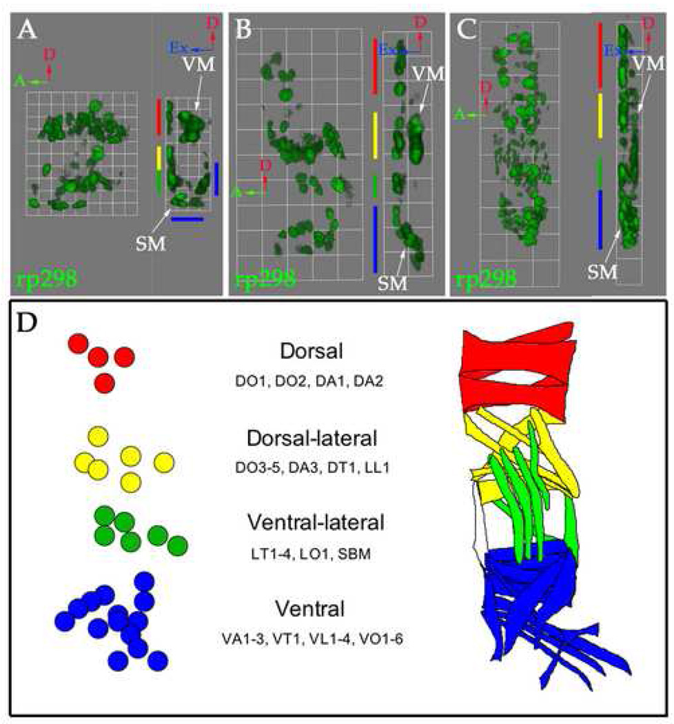
Figure 3. Three-dimensional analysis of FC arrangements shows organization into four groups.
Stage 12 (A), 13 (B) and 14 (C) rp298-lacZ embryos were stained with an antibody against βgal to label FC/myotube nuclei (green). (A–C) Three-dimensional renderings of single mesodermal hemisegments at stage 12 (A, 1 grid unit = 5.7 µm), 13 (B, 1 grid unit = 10.9 µm) and 14 (C, 1 grid unit = 14.1 µm) are shown. Each panel shows an external view (left) and a side view rotated 90° clockwise (right). Red arrows point to dorsal, green arrows point to anterior and blue arrows point to external. SM stands for somatic mesoderm and VM stands for visceral mesoderm. The visceral mesoderm was identified based on position and expression of high levels of F-actin by phalloidin staining (data not shown). During these stages, FCs are organized into four groups in the dorsal (red), dorsal-lateral (yellow), lateral (green) and ventral (blue) somatic mesoderm. At stage 12 (A) the most ventral FCs are located internally. After germband retraction these cells move externally (B–C). Visceral FCs can be clearly seen in the absence of FCMs (put in arrows). These data gathered at each stage have been confirmed with specific FC identity markers. (D) A map of FC arrangements, at stage 13 when 30 FCs can be counted, based on the position and known sibling relationships of the FCs. This map was made by drawing over a flattened version of panel 3B to mark the positions of these cells. A map labeling the identity of each muscle is shown in Supplemental Figure 1.

